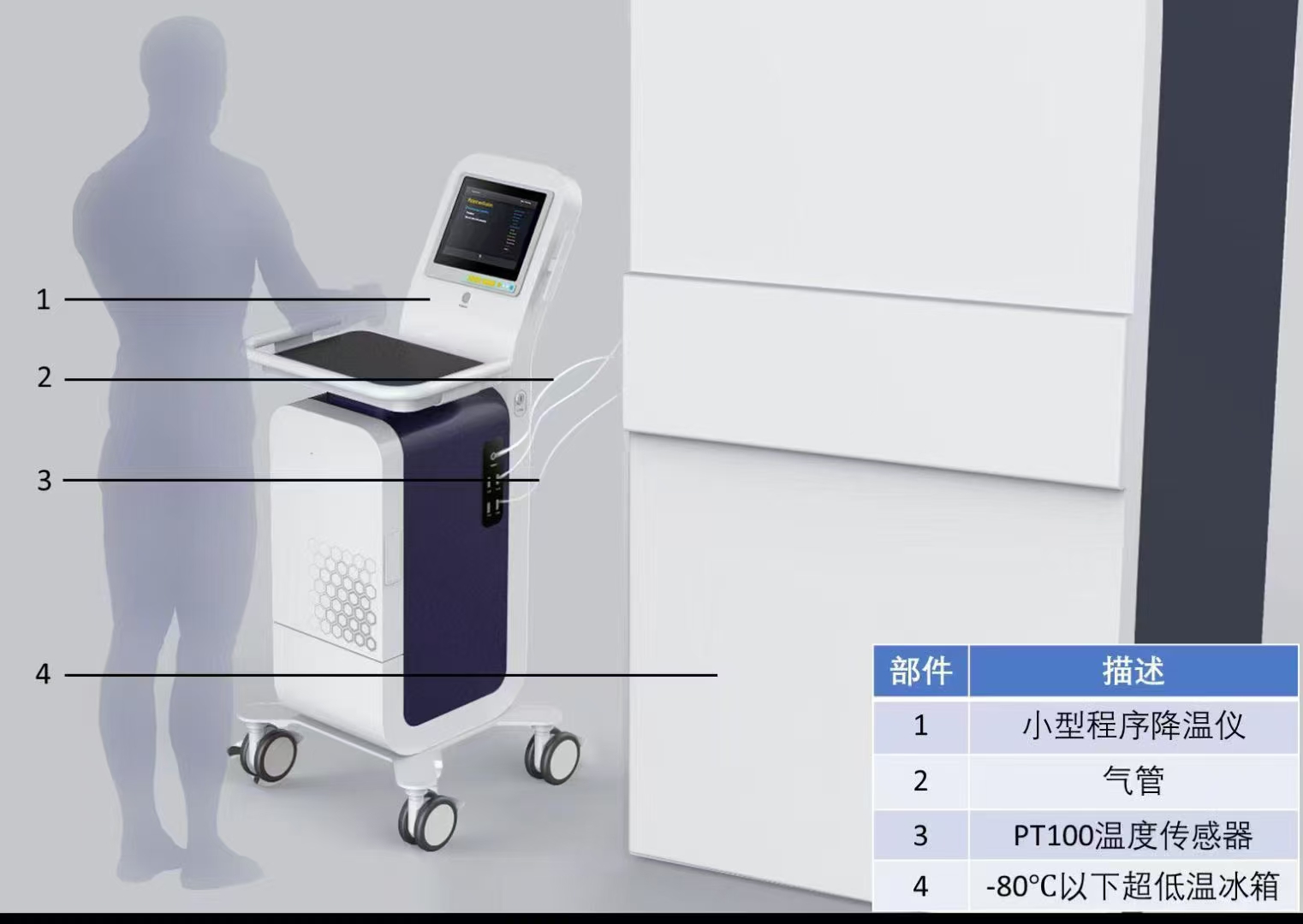
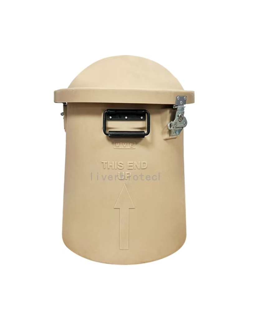
Transport Mode
Liquid Nitrogen Transportation
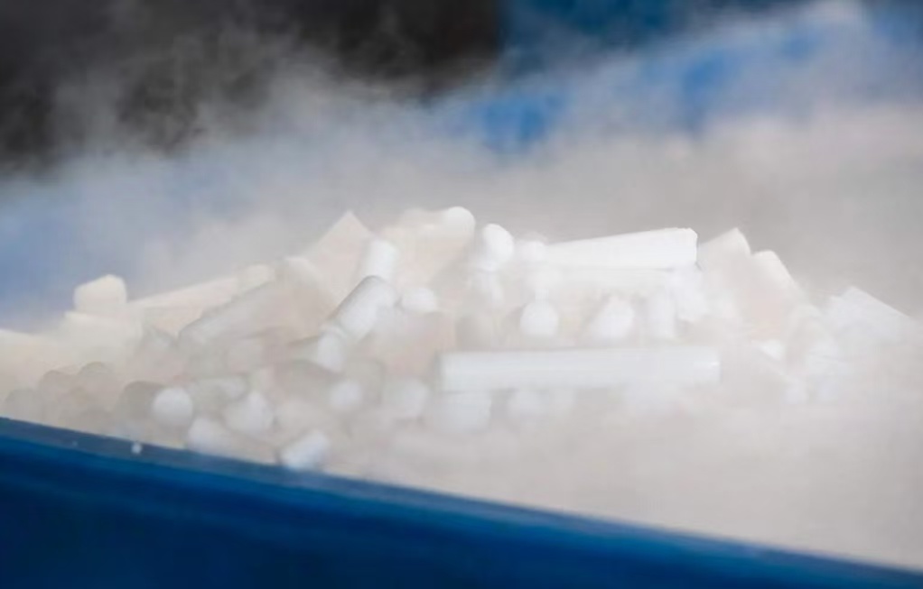
1、Adsorptive Liquid Nitrogen Transportation ,No free liquid nitrogen ,White vapor emission indicates normal operation.
2、Temperature monitoring devices should closely monitor the temperature inside the tank during transportation. If any abnormalities are detected, you can copy the PDF and Excel data from the temperature recorder. (One end of the temperature monitoring device can be unplugged to reveal a USB connector. Simply insert it into a computer to copy the data. If you encounter any difficulties, you can contact the sales staff and technical support for assistance.)
3、Alloy Combination Lock Designed to ensure no third-party access to the LN₂ tank or cell samples during transportation from dispatch to receipt, thereby safeguarding cell integrity.
4、GPS Tracker Enables real-time tracking of cell transportation routes to prevent loss.
5、Liquid Nitrogen Tank, temperature monitoring device, alloy combination lock, and GPS tracker are returnable components. Please store them properly and avoid damage; failure to comply will result in blacklisting.
Reference Citation
pass over.
Scope of Application
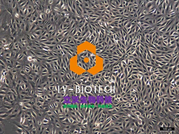
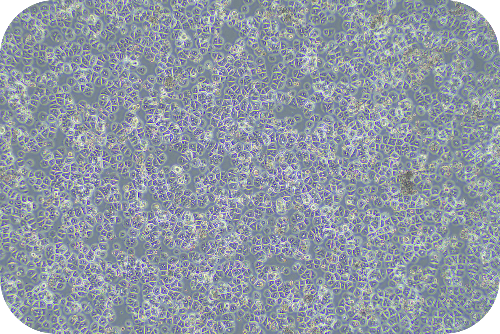
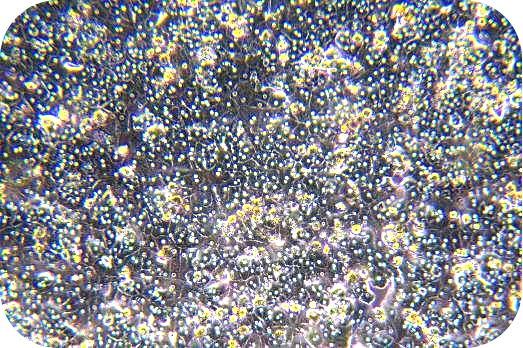
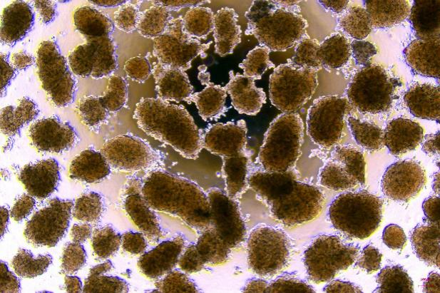
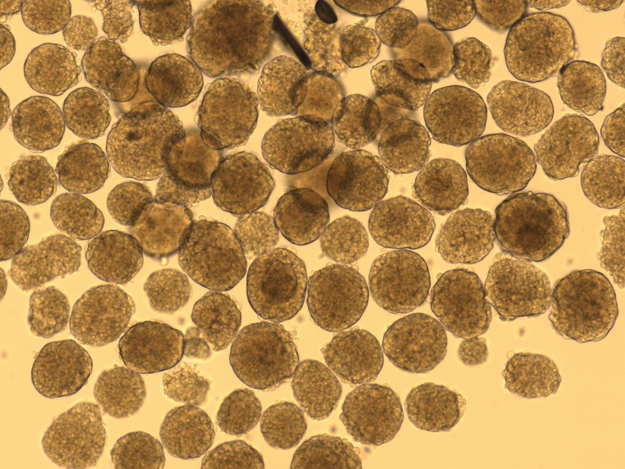
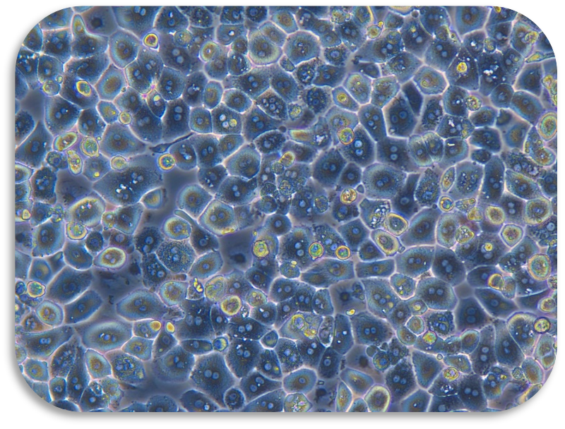
Renal tubular epithelial cells are used to study the mechanisms of acute and chronic kidney disease (such as acute kidney injury, chronic kidney disease), drug nephrotoxicity assessment, renal fibrosis process, cell regeneration and repair mechanism, as well as metabolic disorders (such as diabetic nephropathy) and signal pathway (such as TGF-β, Wnt) regulation, and as a disease model and drug screening platform.
Due to biannual updates of our product instruction manual, the following procedures are for reference only.
The most recent version included with the product shall prevail.
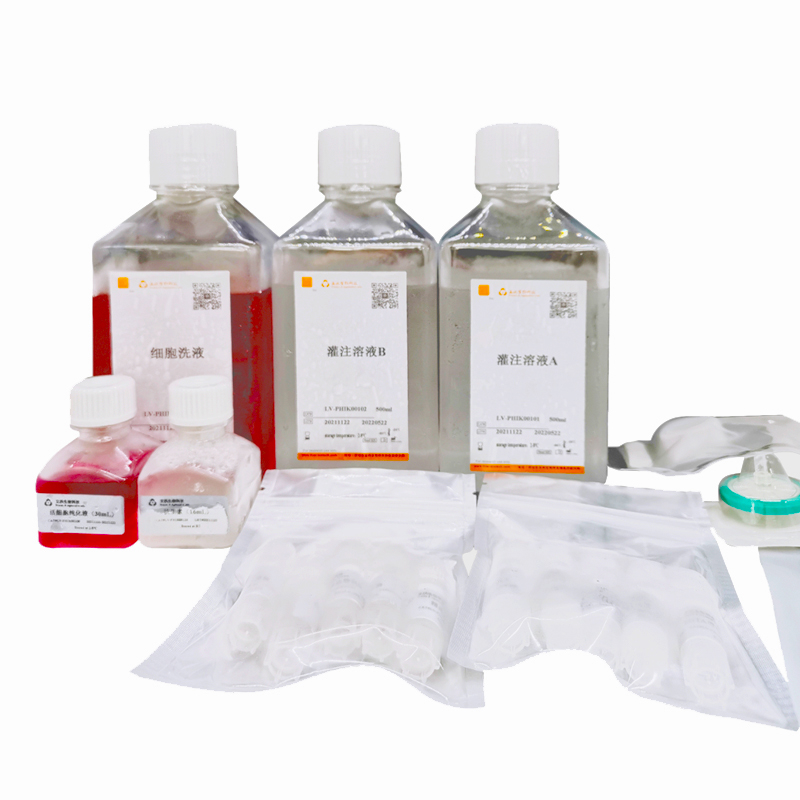
II Reagents and Materials
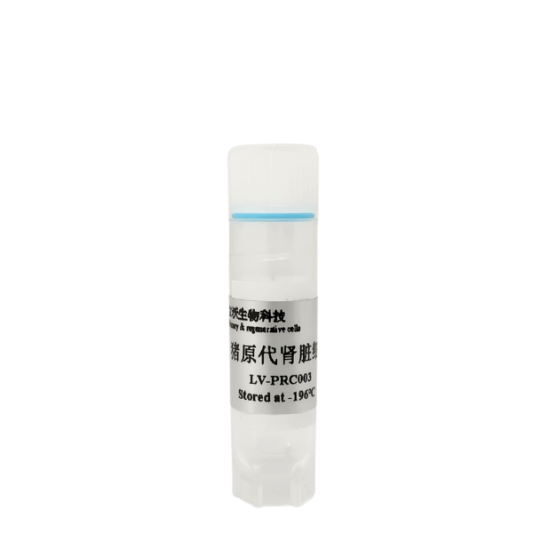
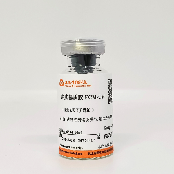
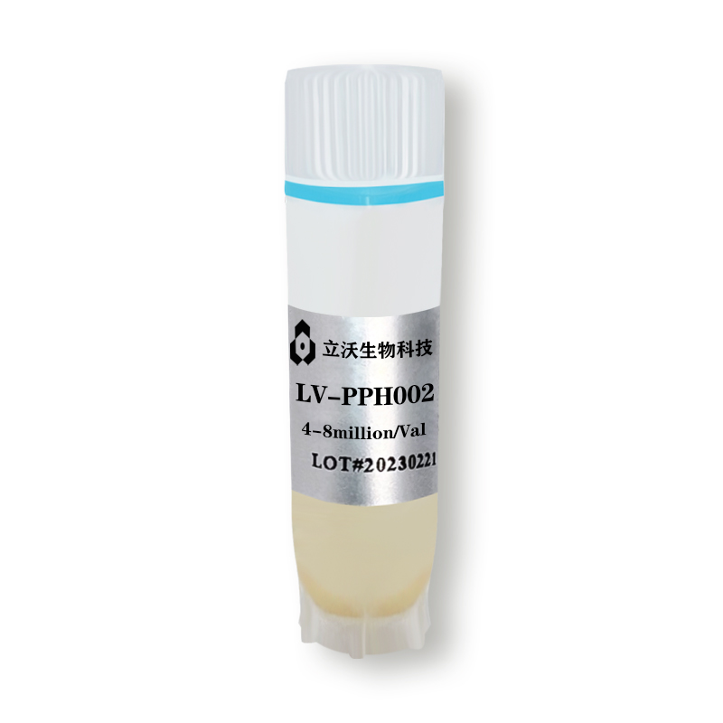
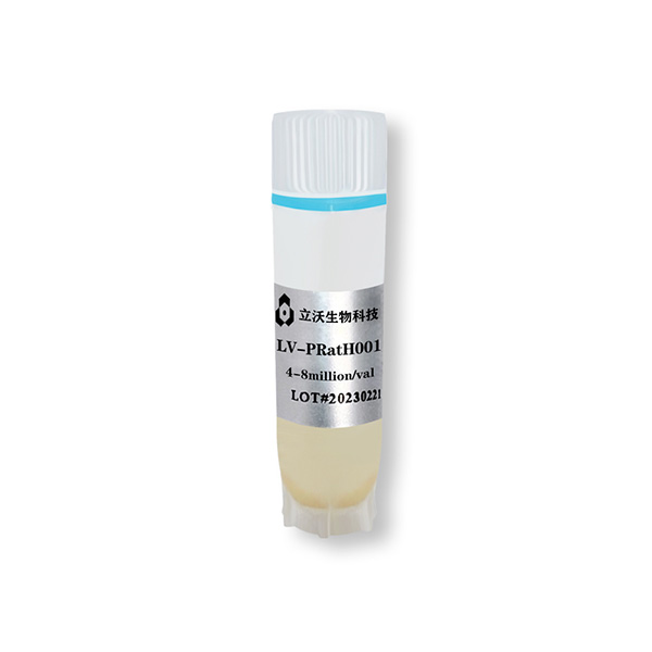
- Frozen renal cortex cell from pig(Cat# LV-PRC001)
- Recovery medium(Cat#LV-Rec001)
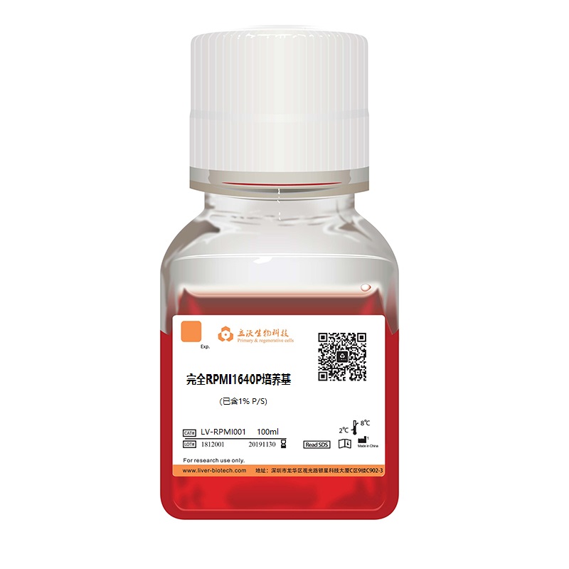
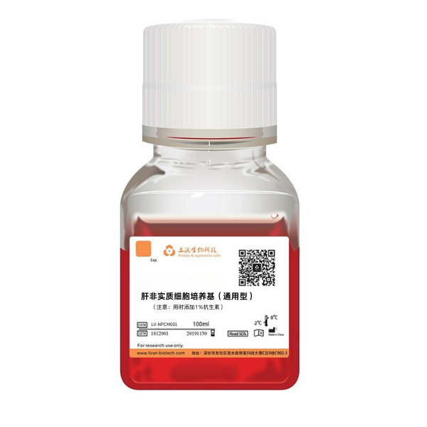
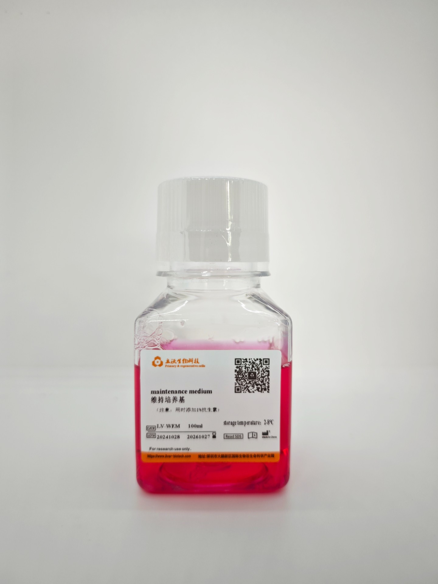
- Renal cortical cell culture medium(Cat# LV-PRCM 001)
- Crushed ice and ice boxes
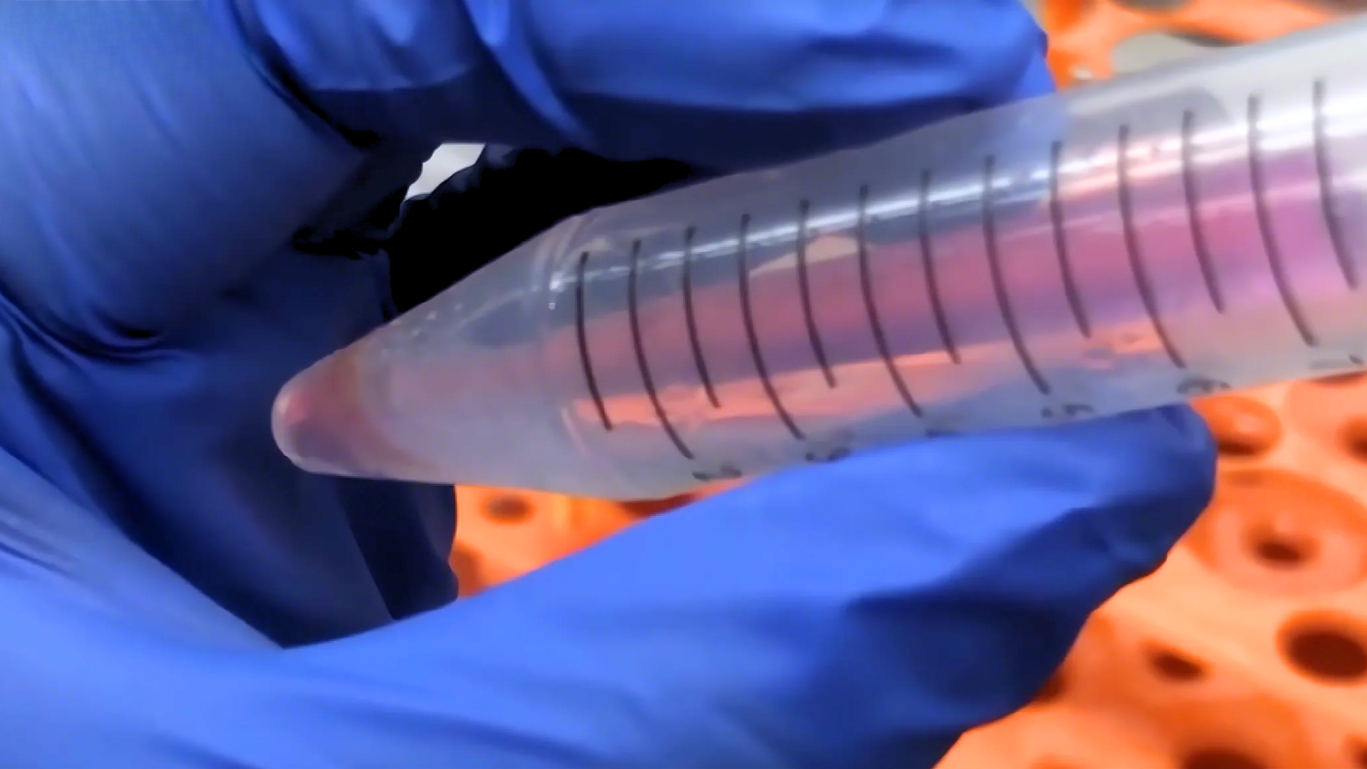
- Sterile centrifuge tube of 15 ml (pre-cooled on ice)
- Disposable pipettes
-Wide-mouth pipette tip (normal pipetting tip is cut off and sterilized)
- Pipet tip
-Thermostat water bath (Preheat at 37°C. Please calibrate the temperature with a thermometer)
- Refrigerated horizontal centrifuge (with horizontal rotor. It is possible to centrifuge 15 ml of centrifuge tubes)
- Biosafety cabinet
- 37 °C/5% CO2 incubator
- 75% alcohol
III Resurgence of Suspension Cells
1. Insert the 15 ml centrifuge tube into an ice box containing enough crushed ice and sterilize it UV for 15 min. Precool centrifuge at 4 °C.
2. Put the recovery medium in crushed ice and pre-cool it well, and put the plating medium into a 38 °C thermostat water bath to fully preheat.
3. Quickly transfer the frozen hepatocytes from the refrigerated position to a thermostat water bath of 38℃, and immerse them in as much water as possible at 38°C. Shake it clockwise and horizontally, but make sure that the cap of the freezing tube is kept above the water.
4. Thaw the freezing tube for about 90-120s, until only a small amount of crushed ice floats in it.
5. Sterilize the freezing tube with 75% alcohol and transfer it to a biosafety cabinet.
6. Resuspend the cells with a wide-mouth pipette tip (gently blow 2 times) and transfer it to a pre-chilled 15 ml centrifuge tube. (Note: if there are cells left on the freezing tube and the pipette tip, 1 ml of recovery medium can be used to clean the them.
7. Add pre-chilled recovery medium to the cell suspension dropwise and10 ml of recovery medium per 1 ml of cell suspension. (Note: the first 3 ml should be added slowly drop by drop and shaken slightly, the next 7 ml can be accelerated). Finally, mix slightly upside down 1 time.
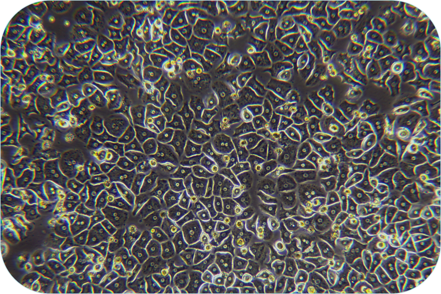
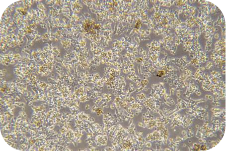
8. Centrifuge of 220 × g at 4 °C for 3 min, remove the supernatant and resuspend with renal cortical cell medium. The survival rate and total number of renal cortical cells can be measured by trypan blue exclusion.
9. Pelleted cells should be resuspended with renal cortical cell medium and settled the volume to 4 ml. Determination of viability and total cell volume of renal cortical cells by trypan blue exclusion.
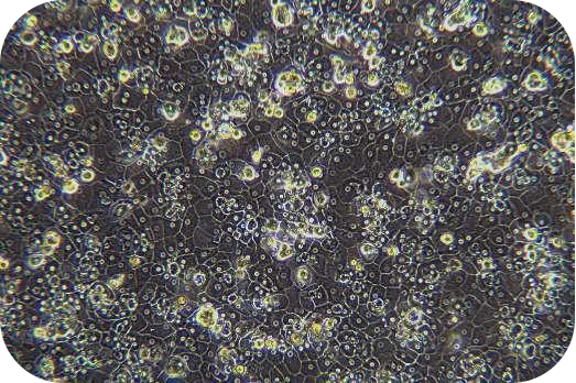
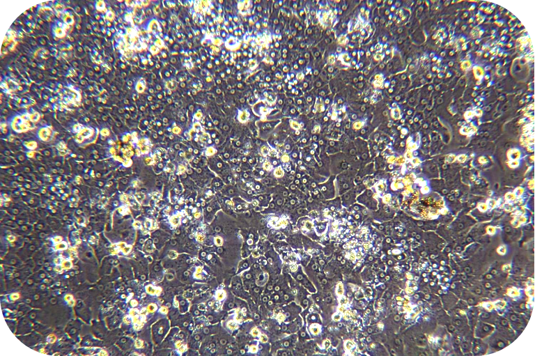
10. Cells can be seeded into a culture dish at a density of 2×10⁴cells/cm2, shaken well and cultured in a 37 °C/5% CO2 incubator. The solution should be changed for 16-24 h.
IV Renal Cortical Cell Culture and Passage
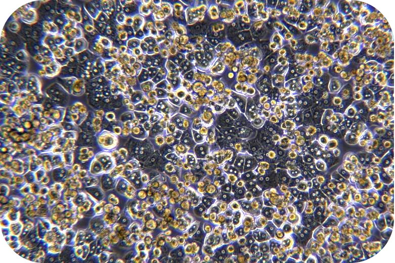
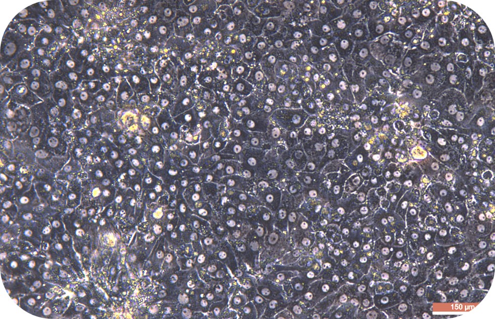
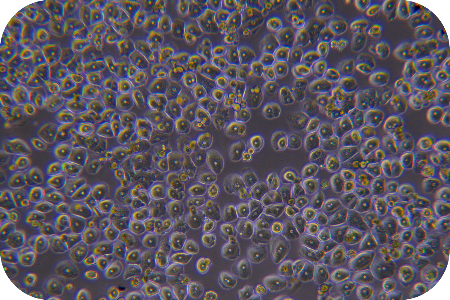
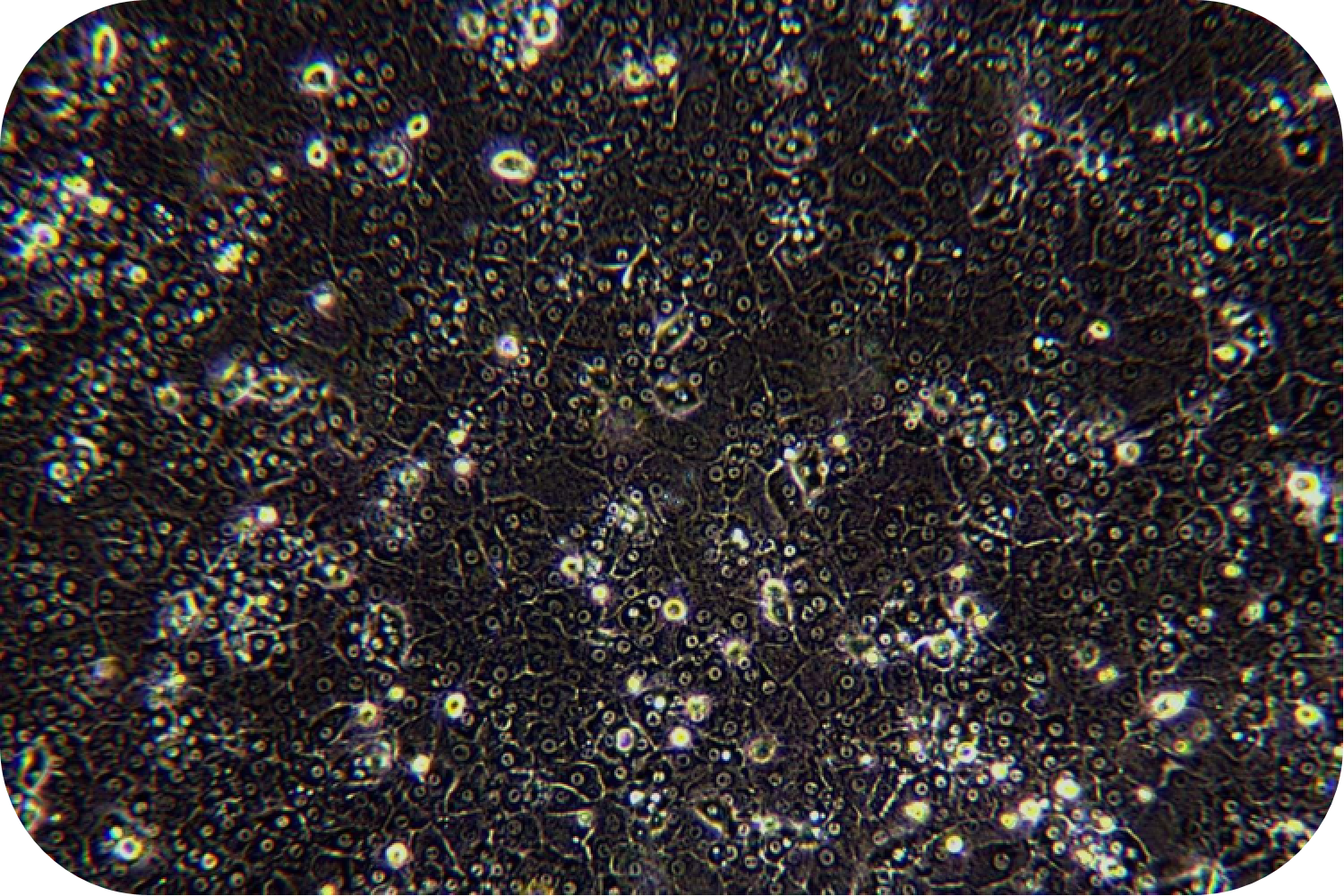
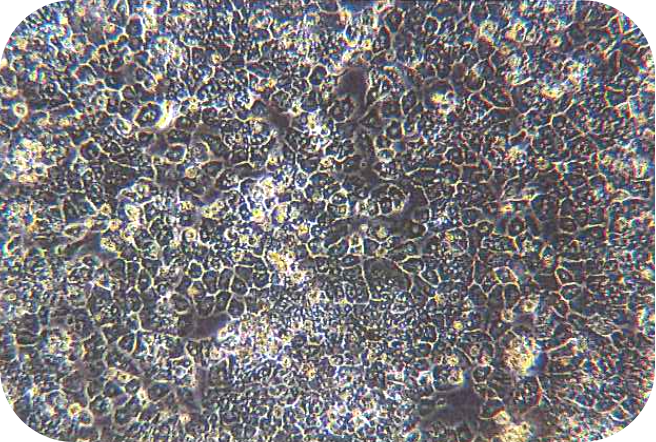
1. The cells can be passaged when the confluency reaches 70-90%.
2. Put the medium, PBS and pancreatin into a 37 °C thermostat water bath to preheat. Wipe with 75% alcohol and then put it into the ultra-clean table.
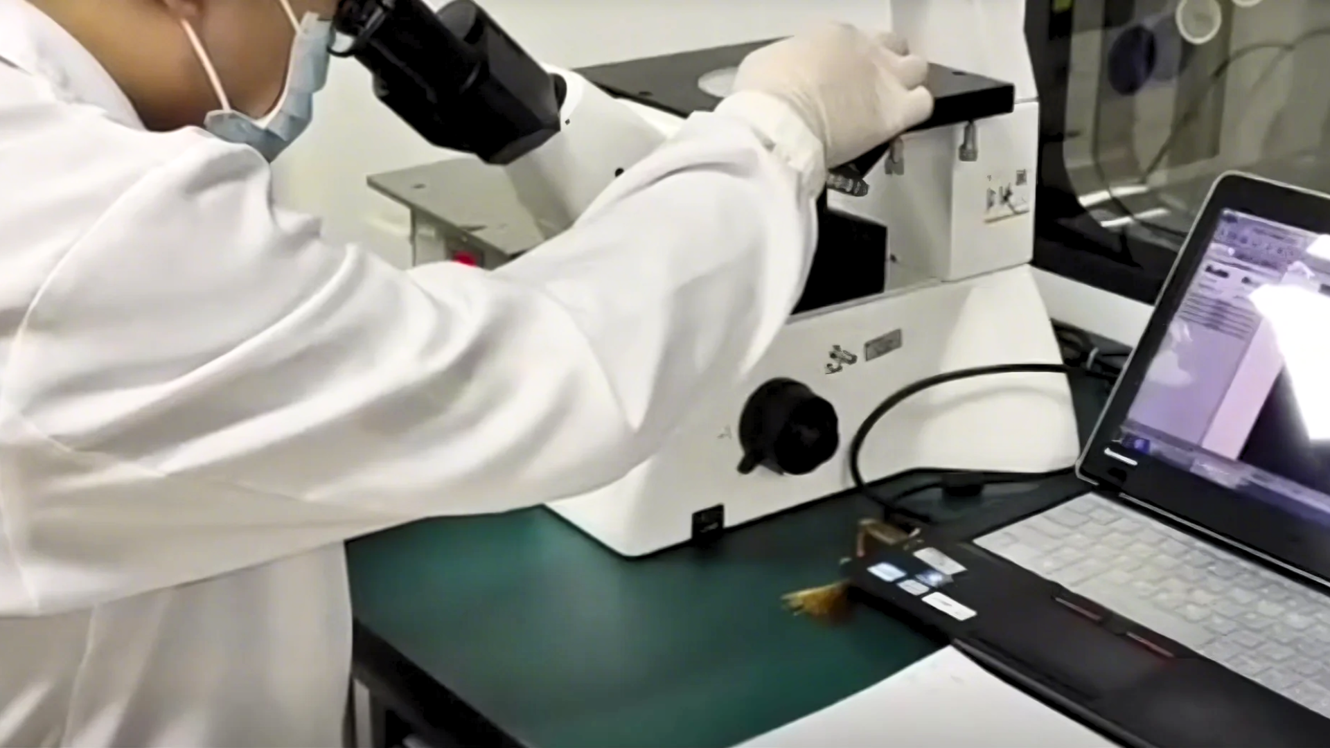
3. Aspirate the old culture solution and add a small amount of PBS to wash the cells. In addition, add an appropriate amount of pancreatin so that the amount of pancreatic enzyme can cover the cells. Incubate the cells at 37°C. Then observe the state under the microscope every 2 to 3 minutes.
4. Discard the pancreatin when the intercellular space of the adherent becomes larger and the cells tend to be rounded but have not yet floated. Add fresh renal cortical cell culture medium and shake the cell flask to stop the action of pancreatin. Carefully blow the adherent cells with a pipette to make a cell suspension (control the force of the blowing to avoid generating a large number of bubbles).
5. Centrifuge at room temperature of 220 × g for 3 min, remove the supernatant and resuspend with renal cortical cell culture medium.
6. Cells can be passaged at a ratio of 1:3 or 1:4. The cell suspension should be seeded into a new cell flask and cultured in a 37 °C/5% CO2 incubator. The growth of the cell adhesion can be observed every other day.
V Customer Service
If you find any quality problems with the product, please collect the original data and contact the company's salesmen or technical support at the first time. The company ensure after-sales service. Every laboratory has different conditions, different operating habits, different proficiency, and objective factors in experimental failure. If the operation is not strictly in accordance with the instructions or exceeds the time limit of after-sales, the company does not do after-sales. Please understand and support us.
Validity period and raw data provided:
Resuscitation problems: Within 24 hours of resuscitation, trypan blue staining or PI staining should be provided.
Pollution problems: Within 96 hours of resuscitation, microscope photographs of differences should be provided.
Purity issues: Within one month, immunofluorescence or flow cytometry results should be provided.
VI Contact Number
Tel:0755-28284050
Technical Support: 19902901483 (Dr. Zhou)