Transport Mode
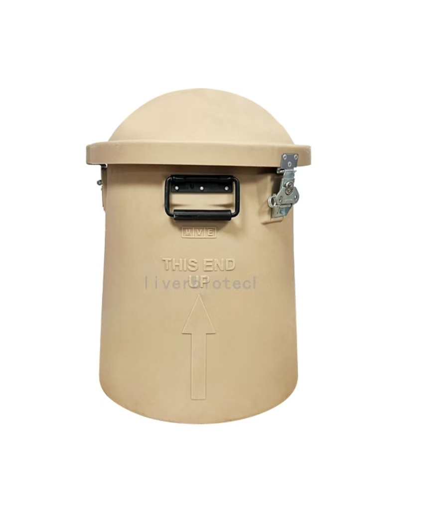
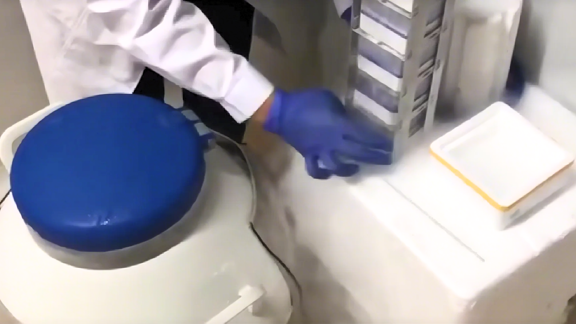
Liquid Nitrogen Transportation
1、Adsorptive Liquid Nitrogen Transportation ,No free liquid nitrogen ,White vapor emission indicates normal operation.
2、Temperature monitoring devices should closely monitor the temperature inside the tank during transportation. If any abnormalities are detected, you can copy the PDF and Excel data from the temperature recorder. (One end of the temperature monitoring device can be unplugged to reveal a USB connector. Simply insert it into a computer to copy the data. If you encounter any difficulties, you can contact the sales staff and technical support for assistance.)
3、Alloy Combination Lock Designed to ensure no third-party access to the LN₂ tank or cell samples during transportation from dispatch to receipt, thereby safeguarding cell integrity.
4、GPS Tracker Enables real-time tracking of cell transportation routes to prevent loss.
5、Liquid Nitrogen Tank, temperature monitoring device, alloy combination lock, and GPS tracker are returnable components. Please store them properly and avoid damage; failure to comply will result in blacklisting.
Reference Citation
pass over.
Scope of Application
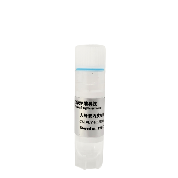
Human liver sinusoidal endothelial cells (LSECs), as crucial non-parenchymal cells in the liver, have become pivotal research subjects in multiple scientific fields due to their unique structural and functional properties. Below is a summary of their primary research applications:
1. Research on Mechanisms of Liver Diseases
LSECs play central roles in multiple hepatic pathological processes, serving as essential models for investigating hepatic fibrosis, fatty liver disease, hepatocellular carcinoma (HCC), and liver regeneration:
Hepatic fibrosis and liver cirrhosis: Defenestration (capillarization) of liver sinusoidal endothelial cells (LSECs) is an early event in hepatic fibrosis, driving the fibrotic process by activating hepatic stellate cells (HSCs) to secrete collagen.
Fatty Liver Diseases: LSECs modulate the pathological progression of non-alcoholic fatty liver disease (NAFLD) and alcoholic fatty liver (AFL) through lipid metabolism regulation (e.g., STAT3 signaling) and anti-inflammatory functions.
Hepatocellular Carcinoma (HCC): LSECs secrete pro-angiogenic factors (e.g., VEGF) to actively participate in tumor microenvironment remodeling and metastatic regulation, while aberrant expression of their signature proteins (e.g., PLVAP) demonstrates prognostic correlation in HCC clinical outcomes.
Liver Regeneration: LSECs synergize with hepatocytes in secreting growth factors (e.g., Hepatocyte Growth Factor, HGF) to orchestrate post-injury tissue repair and functional restoration through spatially-regulated paracrine signaling.
2. Immune Regulation and Tolerance Mechanisms Research
Liver sinusoidal endothelial cells (LSECs) play dual roles in the hepatic immune microenvironment and serve as key targets for studying immune tolerance and inflammatory balance:
Induction of immune tolerance: By expressing scavenger receptors (such as SR-H) and pattern recognition receptors, LSECs clear pathogens and suppress T cell activation to maintain immune tolerance in the liver, but this may also lead to persistent viral infection or tumor immune escape.
Inflammatory regulation: LSECs secrete anti-inflammatory factors (such as prostaglandins, nitric oxide) to suppress excessive inflammatory responses, while clearing endotoxins and inflammatory mediators from the blood through their endocytic function.
Coordination of the entero-hepatic immune axis: LSECs play a bridging role in gut-derived antigen processing by regulating immune cell migration (e.g., macrophages, T cells), thereby influencing the progression of inflammatory liver diseases.
The highly efficient scavenger function of LSECs renders them valuable in drug delivery and toxicity assessment:
Drug clearance mechanism: LSECs rapidly clear macromolecules (e.g., lipoproteins) and nanoparticles from the blood via scavenger receptors (such as Stabilin-1/2), influencing drug half-life and efficacy—key factors in optimizing nanomedicine design.
Targeted therapy development: Investigating the clearance mechanisms of LSECs can provide strategies to reduce non-specific drug uptake, such as modifying the surface of nanoparticles to evade recognition by LSECs.
Toxicity assessment models: The sensitivity of LSECs to drugs or environmental toxins (such as sinusoidal capillarization) is frequently used to evaluate mechanisms of liver injury and screen for protective drugs.
4. Vascular Biology and Microcirculation Research
The unique vascular properties of LSECs provide a research platform for angiogenesis and microcirculation regulation:
Vascular permeability regulation: The fenestral structure and dynamic changes of LSECs (regulated by the Rho-ROCK pathway) influence substance exchange across liver sinusoids, making them an ideal model for studying mechanisms of vascular permeability.
Coagulation and Anticoagulant Balance: Liver sinusoidal endothelial cells (LSECs) maintain sinusoidal blood flow patency by secreting anticoagulant mediators such as heparin sulfate proteoglycans (HSPGs) and prostacyclin (PGI2). Dysregulation of these functions contributes to prothrombotic complications in liver diseases, including portal vein thrombosis and sinusoidal obstruction syndrome.。
Angiogenesis Research: LSECs drive vascular remodeling processes by secreting pro-angiogenic factors (e.g., Vascular Endothelial Growth Factor [VEGF], Angiopoietin-2), playing dual regulatory roles in both physiological liver regeneration and pathological tumor neovascularization.
5. Biomaterials and Tissue Engineering Applications
The unique biological properties of LSECs hold significant potential for advancing artificial liver support system (ALSS) development and innovating next-generation anticoagulant biomaterials.
Artificial liver devices: The endothelialized surface of LSECs mimics the metabolic and immune barrier functions of natural liver sinusoids, serving as a basis for constructing bioreactors or organ-on-a-chip systems.
Anticoagulant materials: The natural anticoagulant properties of LSECs (such as the secretion of PGI₂) are used in designing antithrombotic coatings to improve the biocompatibility of medical devices.
Conclusion
The research applications of human liver sinusoidal endothelial cells (LSECs) span multiple fields, including mechanisms of liver diseases, immune regulation, drug development, vascular biology, and biomaterials. Their multifunctional properties make them a crucial bridge connecting basic research and clinical translation. Future studies could further explore novel mechanisms of LSECs in metabolic regulation (e.g., vitamin A storage) and interorgan interactions (such as the entero-hepatic axis).
Summary
The scientific uses of human liver sinusoidal endothelial cells cover a wide range of fields, including liver disease mechanisms, immune regulation, drug development, vascular biology and biomaterials, and their multifunctional properties make them an important bridge between basic research and clinical translation. Future studies could further explore the novel mechanisms of LSECs in metabolic regulation (e.g., vitamin A storage) and cross-organ interactions (e.g., intestinal-liver axis).
Due to biannual updates of our product instruction manual, the following procedures are for reference only.
The most recent version included with the product shall prevail.
II Reagents and Materials
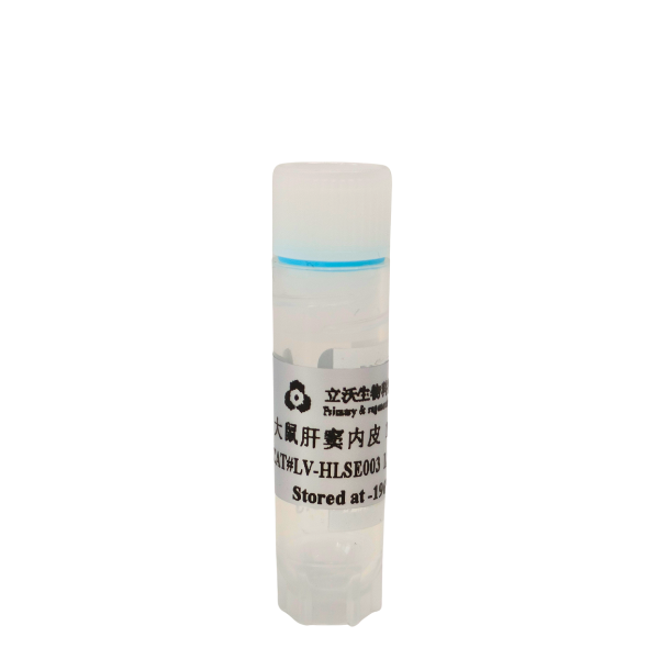
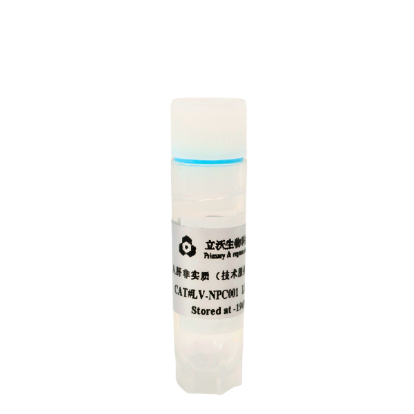
- Rat sinusoidal endothelial cell(Cat# LV-RLSE001)
- Resuscitation medium(Cat#LV-Rec001)
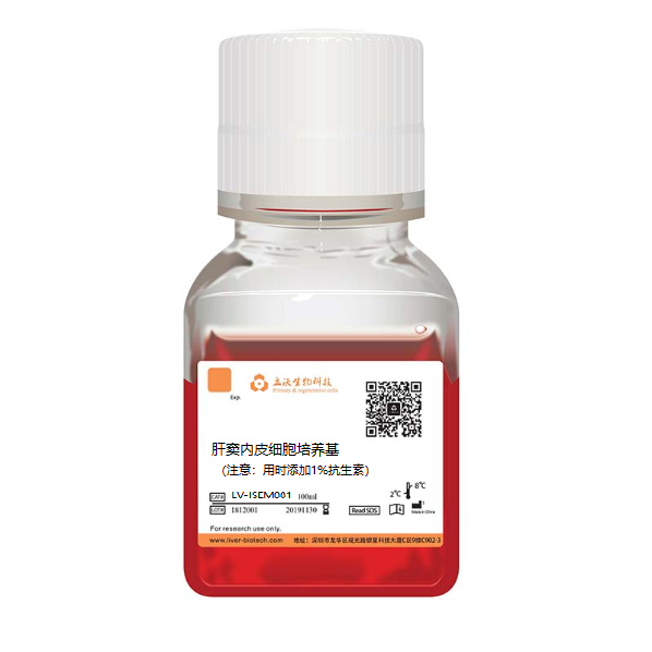
- Culture medium of sinusoidal endothelial cell(Cat# LV-RLSEM001)
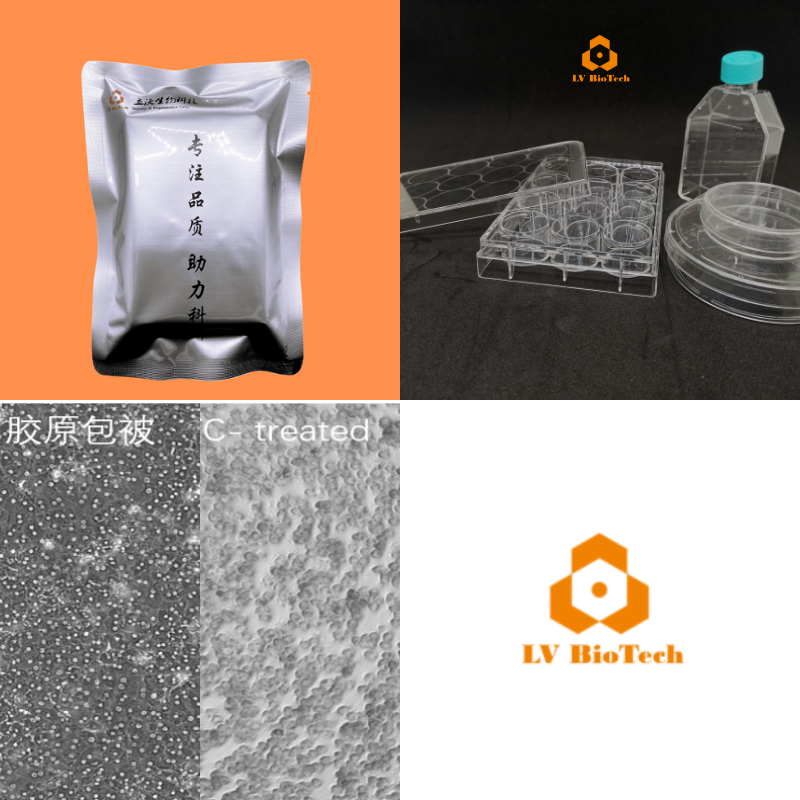
- Collagen-coated plate(LV-coated)
- Crushed ice and ice box
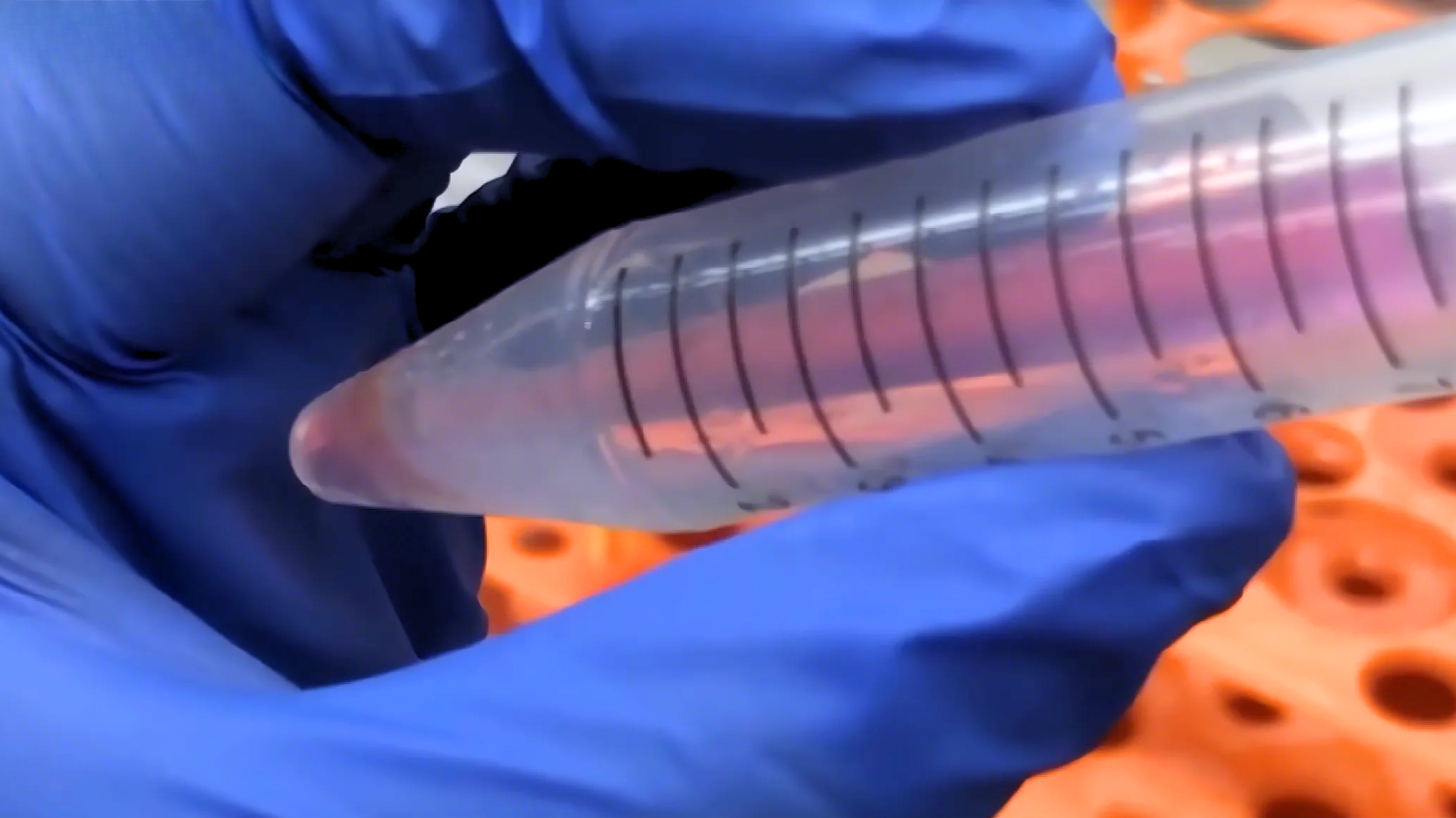
- Sterile centrifuge tube of 15 ml(Pre-chilled on ice)
- Disposable pipette
- Thermostat water bath(Preheat at 38 °C)
- Wide-mouth pipette tip (The tip of a normal one is cut off and sterilized)
-Pipette
- Refrigerated and horizontal centrifuge (with horizontal rotor,it is possible to centrifuge 15 ml of centrifuge tube)
- Biosafety cabinet
-37 °C/5% CO2 Incubator
-75% Alcohol
III Cell Resuscitation and Plating
1. Insert the centrifuge tube of 15 ml into an ice box containing with enough crushed ice and sterilize it UV for 15 min. Then, precool centrifuge at 4 °C.
2. Put the resuscitation medium in crushed ice for fully precooling and put the plating medium into thermostat water bath of 38℃ for fully preheating.
3. Quickly transfer the frozen cells from the refrigerated position to thermostat water bath of 38℃. Immerse them in as much water as possible at 38°C and shake it horizontally and clockwise, but make sure the cap remains above the water.
4. Defrost the freezing tube for about 90-120s, until only a small amount of crushed ice floats in it.
5. Disinfect the freezing tube with 75% alcohol and transfer it to a biosafety cabinet.
6. Resuspend the cells with a wide-mouth tip (gently blow 2 times) and transfer to a pre-chilled freezing tube of 15 ml.
(Note: Residual cells on the freezing tube and pipette tip, they can be washed with 1 ml of resuscitation medium)
7. Add pre-chilled resuscitation medium to the cell suspension dropwise, containing 10 ml of r resuscitation medium per 1 ml of cell suspension (Note: When adding, the first 3 ml should be added slowly dropwise and shaken slightly, and the next 7 ml can be accelerated). Finally, mix slightly upside down once.
8. Centrifuge at 4 °C of 300 × g for 5 min. Remove the supernatant and resuspend with medium.
9. Pelleted cells should be resuspended with the medium of hepatic sinus endothelial cell and settled to the volume of 4 ml. The survival and total number of cells can be measured by trypan blue exclusion.
10. Cells are seeded into collagen-coated plates at 3×10⁴cells/cm2. Shake them well and incubate in a 5% CO2 incubator of 37 °C. After 24 h, change the solution.
IV Cell Culture and Passage
1. The cells can be passaged when the degree of fusion reaches 80%.
2. Put the medium, PBS and pancreatin into thermostat water bath of 37℃ for preheating. And wipe it with 75% alcohol before placing in the ultra-clean table.
3. Aspirate the old culture solution and add a small amount of PBS to wash the cells. In addition, add an appropriate amount of pancreatic enzyme so that the amount of pancreatic enzyme can cover the cells. Incubate the cells at 37°C for 2-3 min.
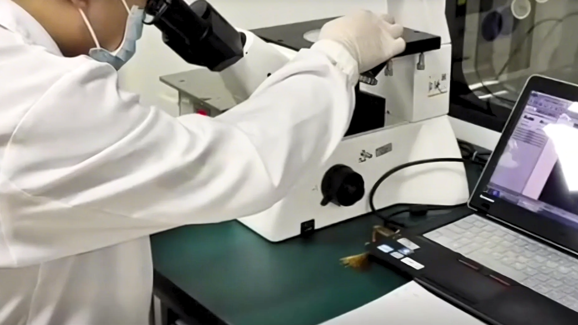
4. Under the microscope, discard the pancreatic enzyme when the intercellular space of the adherent cell becomes larger and the cells tend to be rounded but have not yet floated. After that, fresh hepatic sinus endothelial cell medium should be added and the cell flask should be shaken to terminate the action of pancreatin. Then, carefully blow the adherent cells with a pipette to make a cell suspension (Note:Pay attention to the force of the blowing to avoid generating a large number of bubbles).
5. Centrifuge of 300 × g at room temperature for 5 min. Remove the supernatant and resuspend with medium.
Inoculate the cell suspension into a new collagen-coated flask/plate at the density of 3×10⁴cells/cm2 and place it in a 37 °C/5% CO2 incubator. Adherent growth can be observed every other day.